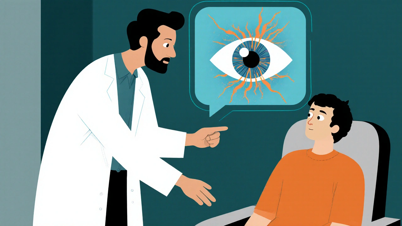
Glaucoma Monitoring: What You Need to Know
When working with glaucoma monitoring, the ongoing process of tracking eye health to prevent vision loss caused by glaucoma. Also known as eye pressure surveillance, it lets doctors adjust treatment before damage becomes permanent.
A core part of glaucoma monitoring is measuring intraocular pressure, the fluid pressure inside the eye that, when too high, can damage the optic nerve. Also called eye pressure, this measurement tells us whether medication needs tweaking. Glaucoma monitoring encompasses intraocular pressure measurement, so regular tonometry appointments are non‑negotiable. Most eye doctors use handheld or table‑top tonometers, and the reading is recorded in mmHg. Keeping a log of these numbers helps spot trends that static snapshots might miss.
The next pillar is optic nerve imaging, techniques like OCT that give a cross‑section view of nerve fibers. Also referred to as retinal nerve fiber layer analysis, this imagery reveals subtle thinning long before a patient notices vision changes. Optic nerve imaging influences treatment decisions in glaucoma monitoring, because it shows whether the disease is progressing despite normal pressure readings. High‑resolution scans are stored digitally, allowing side‑by‑side comparisons over months or years.
Even with pressure under control and a healthy‑looking nerve, functional loss can still creep in. That’s why visual field testing, standardized perimetry that maps a patient’s peripheral vision is a must. Also known as perimetry, it pinpoints blind spots that imaging might miss. Effective glaucoma monitoring requires regular visual field testing, typically every six months for stable cases and more often when changes arise. The results are plotted as a heat map, making it easy for both doctor and patient to see where vision is slipping.
All three measurements feed into one practical goal: maintaining medication adherence and lifestyle habits that protect the optic nerve. Eye‑drop schedules, proper storage, and avoiding activities that spike eye pressure (like heavy lifting) are everyday actions that matter. Some patients benefit from combination drops or laser procedures, but the decision always circles back to the data gathered through pressure checks, imaging, and field tests. When the numbers line up, doctors can confidently keep the current plan; when they diverge, a tweak—whether a new drug, a laser trabeculoplasty, or a surgical option—can be made quickly.
Key Takeaways for Effective Monitoring
In short, glaucoma monitoring is a three‑pronged approach: track intraocular pressure, capture optic nerve health with imaging, and assess functional vision through visual field tests. Each component talks to the others, creating a feedback loop that drives timely treatment changes. Below you’ll find a curated list of articles that dive deeper into each tool, explain how to interpret results, and offer tips on staying on top of your eye health. Use them to build a personalized monitoring routine that keeps your sight safe for the long haul.
Glaucoma and Visual Field Testing: What to Expect
Learn what to expect during a glaucoma visual field test, why it matters, how it works, preparation tips, result interpretation, and next steps for managing your eye health.