
Living with an autoimmune disease isn’t just about taking medication. It’s about staying ahead of your body’s signals-before the fatigue hits, before the joint pain becomes unbearable, before organ damage sets in. That’s where autoimmune disease monitoring comes in. It’s not a one-time test. It’s a system. A routine. A way to catch trouble early, track how well treatment is working, and avoid hospital visits when you can prevent them.
What Lab Tests Actually Tell You
Lab work is the foundation. But not all blood tests are created equal. Many people think a positive ANA test means they have lupus. It doesn’t. About 20% of healthy people test positive for ANA. That’s why it’s a screening tool, not a diagnosis. If your ANA is positive, doctors don’t stop there. They look at reflex tests-like anti-dsDNA, SS-A, SS-B, Scl-70, and Jo-1. These are the real clues.
Anti-dsDNA is specific. If it’s high, especially in someone with lupus, it often means the kidneys are under stress. A rising level can warn of lupus nephritis before you even feel symptoms. Complement levels (C3 and C4) are just as important. When they drop, inflammation is active. Many patients get stuck in a loop of checking ANA every visit. That’s a waste. ANA stays positive even when you’re in remission. Complement levels? Those move with your disease.
C-reactive protein (CRP) and erythrocyte sedimentation rate (ESR) measure inflammation. CRP above 3.0 mg/L? That’s active inflammation. ESR over 20 mm/hr for women or 15 mm/hr for men? Same thing. But here’s the catch: these markers aren’t perfect. In some autoimmune diseases like lupus, CRP can stay normal even when there’s major flare. That’s why you need the full picture-antibodies, complements, and clinical symptoms together.
Imaging: Seeing What Blood Can’t Show
Lab tests show what’s happening inside your blood. Imaging shows what’s happening inside your joints, organs, and tissues. MRI is the gold standard for spotting early inflammation. It can see swelling in the synovium (joint lining) long before X-rays show bone damage. Newer MRI techniques with nanotech contrast agents are safer and more precise than old gadolinium ones, which carried risks of kidney damage.
Ultrasound is underrated. It’s fast, cheap, and doesn’t use radiation. With microbubble contrast, it can measure blood flow in inflamed joints. In rheumatoid arthritis, it’s 85% accurate at detecting active disease-better than some blood markers. For patients with vasculitis or muscle inflammation, ultrasound can pinpoint exactly where the problem is.
PET scans are changing the game. By tagging immune cells with radioactive tracers, doctors can now see where T-cells are congregating in the body. This isn’t just for cancer anymore. In autoimmune conditions, it’s revealing hidden pockets of inflammation that labs miss. SPECT scans do something similar, using peptides that bind to inflammation sites to create 3D maps of immune activity.
CT scans are used less often because of radiation exposure, but they’re still vital when checking for lung scarring in scleroderma or bowel damage in Crohn’s disease. The key? Use imaging not just to confirm damage, but to catch it before it’s permanent.
How Often Should You See Your Doctor?
There’s no one-size-fits-all schedule. It depends on your disease, how active it is, and how you’re responding to treatment.
When you’re newly diagnosed or starting a new drug, expect visits every 4 to 6 weeks. That’s when your doctor needs to check for side effects, adjust doses, and see if the medication is working. Once your disease is under control, visits usually stretch to every 3 to 4 months. The American College of Rheumatology says you should still have at least two full assessments per year-even if you feel fine.
For stable disease, some patients can go 6 to 12 months between visits, especially if they’re on maintenance therapy and have no organ involvement. But if you’ve had kidney, lung, or heart complications in the past, quarterly visits are non-negotiable. The European League Against Rheumatism (EULAR) recommends monthly visits during active flares. That’s not overkill-it’s prevention.
Don’t wait for a flare to show up. Track your symptoms. Keep a journal. Note morning stiffness, sleep quality, rashes, or new swelling. Bring it to your visit. Your doctor can’t adjust your treatment if they don’t know what you’re experiencing.
The 3-Pillar Approach to Monitoring
Experts agree: the best monitoring uses three pillars-labs, imaging, and clinical assessment. And each one carries weight.
At the 2023 International Autoimmune Summit, doctors from across the globe agreed on this breakdown: 30% lab markers, 30% imaging findings, and 40% clinical evaluation. Why does clinical matter more? Because symptoms don’t always match the numbers. A patient might have normal CRP and ESR, but still be in pain, exhausted, and unable to work. That’s a flare. And if you’re only looking at labs, you’ll miss it.
Dr. Betty Hahn from UNC put it simply: “Relying on labs alone misses critical context in 63% of flares.” That’s not a small number. That’s the majority.
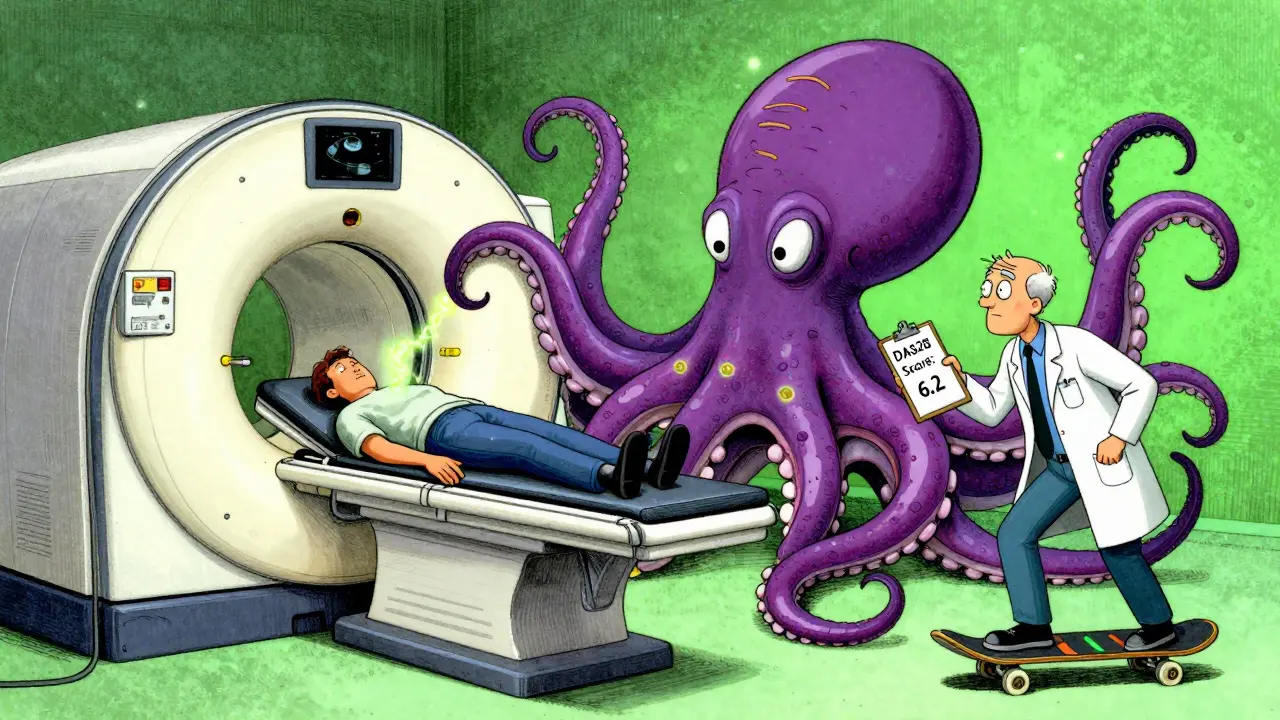
Doctors now use disease activity scores to track progress. For rheumatoid arthritis, it’s DAS28. For lupus, it’s SLEDAI. These scores combine joint counts, lab values, and patient reports into one number. They help decide if you need to switch drugs or if you’re doing well enough to stay on current treatment.
What’s New in Monitoring
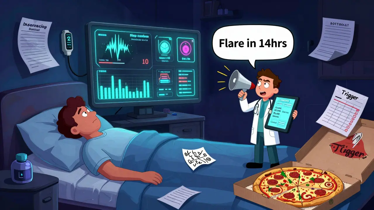
Technology is moving fast. Wearables are no longer just for counting steps. New devices can analyze interstitial fluid-fluid between your cells-for markers like CRP and cytokines. Early studies show 89% correlation with traditional blood tests. Imagine getting a daily alert on your phone: “Your inflammation is rising. Consider resting today.” That’s not science fiction anymore.
AI is stepping in too. Algorithms now analyze years of your lab results, imaging reports, and symptom logs to predict flares. One system, tested in 2,347 patients, predicted flare-ups 14 days in advance with 76% accuracy. That’s enough time to call your doctor, adjust your steroid dose, or skip a stressful event.
The FDA approved the first integrated platform called AutoimmuneTrack in mid-2023. It pulls data from wearables, lab systems, and patient apps into one dashboard. In a year-long trial, users had 29% fewer emergency visits. That’s huge.
Barriers and Real-World Challenges
Not everyone has access to this level of care. Insurance often won’t cover frequent MRIs or PET scans. Only 48% of Medicaid patients get the recommended monitoring, compared to 83% of those with private insurance. That’s not just unfair-it’s dangerous. Delayed monitoring means more organ damage, more hospitalizations, more disability.
Another problem? Test variability. ANA results can differ by 22% between labs. One place says “positive,” another says “equivocal.” That’s why it’s critical to stick with the same lab and same testing method when tracking over time.
And while CyTOF (mass cytometry) lets scientists analyze 50 immune cell markers at once, it’s still mostly in research labs. It’s expensive, complex, and not yet routine in clinics. But it’s coming. The market for autoimmune monitoring is growing fast-$12.7 billion globally-and that means better tools will become more accessible.
What You Can Do Today
You don’t need the latest tech to take control. Start here:
- Know your baseline numbers. Get copies of your last CRP, ESR, ANA, anti-dsDNA, and complement levels.
- Ask your doctor: “Which tests should I track every visit? Which ones are just for diagnosis?”
- Keep a simple symptom journal. Note pain levels, energy, sleep, swelling, and new rashes.
- Don’t skip imaging if recommended. Even one MRI or ultrasound can show hidden damage.
- Push back if your insurance denies a test. Appeal. Ask for a letter of medical necessity.
- Find out if your clinic uses disease activity scores like DAS28 or SLEDAI. If not, ask why.
Autoimmune disease isn’t something you cure. It’s something you manage. And the best way to manage it? Stay ahead of it. Use every tool available. Labs, imaging, and visits aren’t chores-they’re your early warning system. The sooner you catch a flare, the less damage it does. And the more control you have over your life.
Is ANA testing useful for monitoring autoimmune disease activity?
No, ANA testing is not useful for monitoring disease activity. Once positive, ANA often stays positive even during remission. It’s a screening tool for diagnosis, not a tracker of flare-ups. More useful markers include anti-dsDNA antibodies and complement levels (C3 and C4), which drop when lupus is active. Relying on ANA alone can give false reassurance.
How often should I get imaging for my autoimmune disease?
There’s no fixed schedule. If you have joint disease like rheumatoid arthritis, ultrasound or MRI may be done every 6-12 months to check for early damage. For conditions like lupus or vasculitis, imaging (like chest CT or kidney ultrasound) is done if symptoms suggest organ involvement. Routine imaging isn’t needed if you’re stable and symptom-free. Always follow your doctor’s advice based on your specific disease and risk factors.
Can wearable devices replace lab tests for autoimmune monitoring?
Not yet, but they’re becoming valuable supplements. Wearables that measure inflammatory markers through interstitial fluid show strong correlation (89%) with traditional CRP blood tests. They’re great for spotting trends and early warning signs between visits. But they can’t replace antibody panels, complement levels, or imaging. Think of them as an early alert system, not a full diagnostic tool.
Why do some doctors order so many blood tests?
Some tests are for diagnosis, others for monitoring. Doctors often order a broad panel early on to rule out multiple conditions. Once the diagnosis is clear, they should focus on key markers like CRP, ESR, anti-dsDNA, C3/C4, and disease-specific antibodies. Ordering unnecessary tests like repeat ANA or broad autoimmune panels every visit adds cost and confusion. Ask your doctor which tests are actually helping track your condition.
What should I bring to my autoimmune disease visit?
Bring your symptom journal, a list of current medications (including supplements), recent lab results (if you have them), and any imaging reports. Note changes in energy, pain, swelling, or new symptoms. If you’ve had a recent illness, stress, or change in routine, mention it-those can trigger flares. The more context you give, the better your doctor can interpret your lab and imaging results.
Are there affordable alternatives if I can’t afford frequent imaging?
Yes. Ultrasound is often more affordable than MRI and can be just as useful for joint and muscle monitoring. Ask if your clinic offers ultrasound as part of routine visits. Also, focus on tracking symptoms and blood markers closely-these can often signal when imaging is truly needed. Some patient assistance programs or university hospitals offer reduced-cost scans. Don’t assume you can’t get it-ask.
Comments
Carolyn Benson
December 17, 2025 AT 18:13ANA is a red herring. I’ve been told my ANA is positive for 12 years. Twelve. And I’ve never had a flare because of it. My complement levels? Those are the ones that spike when I’m about to crash. But no one listens. They just keep ordering the same damn test like it’s a horoscope. You don’t monitor a wildfire by checking if the sky is cloudy-you check the heat signature. Stop wasting time and money on ANA.
Aadil Munshi
December 19, 2025 AT 14:12Bro, you’re 100% right about ANA being useless for monitoring-but let’s be real, the whole system is broken. I got my first MRI at 28 because my CRP was high. Took 8 months to get it approved. Insurance said ‘no evidence of structural damage.’ But I couldn’t lift my coffee cup. They don’t care about the invisible stuff until you’re on a gurney. And don’t even get me started on how labs vary by 22%. One lab says ‘positive,’ another says ‘maybe.’ How is that science?
Also, AI predicting flares 14 days out? That’s wild. But who’s gonna pay for it? My Medicaid plan won’t cover a Fitbit that tracks cytokines. Meanwhile, rich people get wearables and PET scans. This isn’t medicine. It’s a luxury subscription.
Frank Drewery
December 20, 2025 AT 10:10This is such a helpful breakdown. I’ve been struggling to explain to my doctor why I feel awful even when my labs look ‘fine.’ The 30-30-40 rule makes so much sense. I started keeping a symptom journal last month-just a simple note every night: ‘pain 6/10, slept 4 hours, swollen knuckles.’ My DAS28 score went down 2 points in 6 weeks just because we had actual data to work with. Small things matter. Thank you for writing this.
Danielle Stewart
December 21, 2025 AT 18:24For anyone reading this: if you’re on maintenance therapy and feel stable, don’t skip your visits. I did. I thought I was fine. Three months later, I had kidney inflammation. No symptoms. Just a drop in C3. That’s it. I was lucky. Others aren’t. Your doctor isn’t just checking boxes-they’re trying to keep you off dialysis. Trust the process. Even if you feel great. Especially then.
mary lizardo
December 23, 2025 AT 18:02Let’s address the elephant in the room: the author’s conflation of ‘monitoring’ with ‘medical surveillance.’ Monitoring implies agency. Surveillance implies control. Who is controlling whom here? The patient? Or the pharmaceutical-industrial complex that profits from endless testing? The $12.7 billion market figure is not a triumph-it’s a red flag. We are being monetized under the guise of ‘early detection.’ And don’t get me started on ‘AutoimmuneTrack’-it sounds like a Silicon Valley buzzword dressed in lab coats.
Furthermore, the claim that ‘symptoms don’t always match the numbers’ is not a revelation-it’s a failure of reductionist medicine. The body is not a spreadsheet. You cannot reduce chronic illness to a series of biomarkers and algorithms. The real crisis isn’t lack of data-it’s the erosion of patient autonomy in favor of quantified control.
Adrienne Dagg
December 24, 2025 AT 14:25OMG YES. I’ve been screaming this for years. ANA is NOT a flare meter. 🙄 My doc kept ordering it every visit until I printed out the 2023 summit paper and shoved it in his face. Now he uses SLEDAI and tracks C3/C4. Also, ultrasound for joints? GAME CHANGER. I got mine at a free clinic-no insurance needed. You don’t need fancy tech to be proactive. Just be loud. And stop trusting ANA like it’s a tarot card. 💪
Glen Arreglo
December 25, 2025 AT 00:49I’ve been in this community for 15 years. I’ve seen people get denied care because their CRP was ‘normal’ while their hands were deforming. I’ve seen doctors dismiss fatigue as ‘stress.’ This post nails it: it’s not about the numbers. It’s about the person behind them. If you’re exhausted, in pain, and your doctor says ‘your labs are fine,’ walk out. Find someone who listens. Your life depends on it.
shivam seo
December 25, 2025 AT 14:10USA medical system is a joke. I came here from Australia and I couldn’t believe how much they charge for an ultrasound. We get them for free here. And PET scans? We use them for autoimmune monitoring too-but only when clinically indicated. Not every 3 months like some kind of ritual. Here, they just pump you full of tests to make money. And then they wonder why people can’t afford care. Pathetic.
benchidelle rivera
December 25, 2025 AT 21:40As someone who’s managed lupus for 21 years, I can tell you this: the most dangerous thing you can do is wait for symptoms to be ‘bad enough’ to warrant action. I lost 30% of my kidney function before I learned to track my own C3/C4 levels. Now I keep a spreadsheet. I bring it to every appointment. My rheumatologist says I’m the most prepared patient she’s ever had. Don’t wait for the system to protect you. Protect yourself. Knowledge is power-and it’s free.
Andrew Kelly
December 27, 2025 AT 05:54Let’s be honest-this whole ‘autoimmune monitoring’ thing is a scam. The FDA approved ‘AutoimmuneTrack’? That’s a product of Big Pharma’s lobbying arm. Wearables measuring cytokines? They’ve been trying to sell this since 2015. And now suddenly it’s ‘revolutionary’? Meanwhile, the real cause of autoimmune disease-glyphosate, 5G, vaccines, EMF-isn’t even being discussed. They want you to think it’s all about ‘labs and imaging’ so you don’t look at the real enemy: the government and the corporations poisoning you. Wake up.
Isabel Rábago
December 28, 2025 AT 04:26ANA is not a diagnostic tool. It is not a monitoring tool. It is not a predictor. It is a screening tool. That is all. Repeating it is medical malpractice. If your physician continues to order it, you are being mistreated. Period. This is not opinion. This is evidence-based medicine. Your time, your money, your body-do not waste them on obsolete, misleading, statistically irrelevant tests. Demand better.
bhushan telavane
December 28, 2025 AT 14:12Hey, I’m from India. We don’t have access to half of this stuff. But I do track my symptoms. Every morning, I write down: pain, fatigue, swelling. I take pics of my hands. I show them to my doctor. He doesn’t have MRI or PET, but he listens. That’s what matters. You don’t need fancy tech to be smart. Just be consistent. And don’t let anyone make you feel like your pain isn’t real because your CRP is ‘normal.’
Mahammad Muradov
December 30, 2025 AT 10:38AI predicting flares? That’s cute. But AI doesn’t live in your body. It doesn’t feel the stiffness in your knees at 4 a.m. or the way your lungs tighten when you climb stairs. You can’t replace lived experience with algorithms. The real innovation is patient advocacy. Not apps. Not wearables. Talking. Asking. Pushing back. That’s the only thing that saves lives in this broken system.
Frank Drewery
December 31, 2025 AT 13:39Just wanted to reply to @6024-you’re so right. I used to think tech would fix everything. Then I realized my doctor didn’t even look at my symptom journal until I printed it out and handed it to him. No app, no AI, no fancy dashboard. Just me, paper, and the courage to say, ‘This is what I’m going through.’ That’s the real breakthrough.